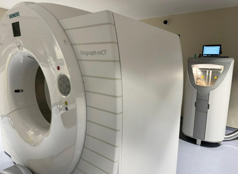
Myocardial perfusion imaging of patients with suspected coronary artery disease is on the cusp of a new era. UVA Health is one of only a handful of health systems in the U.S. gearing up to measure blood flow through myocardial perfusion imaging with O15-water positron emission tomography (PET). This will help clinicians at UVA Health make quicker and more accurate diagnoses of coronary artery disease.
O15-water is a radionuclide tracer. It can help assess, visualize, and measure blood flow to the heart with a high degree of accuracy. This helps diagnose conditions like myocardial ischemia and myocardial infarction.
It has several research and clinical applications, including cardiovascular stress testing.
“We’re very excited to bring this capability to UVA Health because this tracer can be used in any area where we need to assess myocardial blood flow and myocardial scar,” says Jamieson Bourque, MD, UVA Health’s medical director of nuclear cardiology and an Executive Committee member of the American Society of Nuclear Cardiology. Applications include using it for inflammatory diseases like cardiac sarcoidosis, cardiac allograft vasculopathy, and other conditions where blood flow assessment is critical.
Several Research Benefits
O15-water PET imaging has been used in Europe for research and clinical purposes for many years. But it hasn’t yet been approved for clinical use in the U.S.
While UVA Health uses this radionuclide tracer exclusively for research currently, Bourque anticipates eventual clinical approval. UVA Health is participating in a pivotal clinical trial to support this process. “The research applications of the O15-water agent are profound,” says Bourque. “It’s going to help answer advanced questions that have long stymied researchers. At UVA Health, we’re particularly interested in how O15-water can support our research optimizing diagnosis and therapeutic options for patients with microvascular disease.” Likewise, “With cardiac sarcoidosis, we’re studying how the radionuclide tracer can help us observe myocardial scarring and the impact of therapy. Accurate measurement of myocardial blood flow could allow more precise assessment of cardiac allograft vasculopathy post-cardiac transplant,” adds Bourque.
Patient Benefits: Quicker Testing, Less Exposure
The tracer has a very short half-life (about 2 minutes). This short period reduces patients’ exposure to radiation.
Because it dissipates so quickly, labs conducting cardiac stress tests can do faster repeat imaging for rest and stress. This not only increases lab efficiency, but also enables patients to complete the stress test process far more quickly compared with other myocardial perfusion tracers, improving patient satisfaction.
“The entire stress test process can typically be completed in less than 1 hour using the O15-water agent,” says Bourque.
Use of the tracer can also benefit patients with other conditions. “It can be applied broadly to disease states beyond cardiovascular disease,” says Bourque, “including in neurology, urology, and hematology-oncology.”
Creating the Tracer
To produce the O15-water agent, sophisticated equipment must be onsite. Using a cyclotron, which is similar to a proton accelerator, magnetic fields bombard oxygen with high energy protons, thereby creating radioactive oxygen gas.
This gas is then transferred via a pipe into the PET scanner room. There, another machine converts the gas into water, which in turn, is injected into the patient.
“UVA Health was a natural partner to pioneer this perfusion tracer because it’s one of the very few facilities in the nation that has a state-of-the-art PET scanner immediately-adjacent to a cyclotron,” notes Bourque.
Potential Clinical Advantages of O15-Water PET
Myocardial perfusion imaging with O15-water PET improves diagnostic accuracy for assessing obstructive coronary artery disease and the clinical significance of any blockages.
Other potential benefits include the ability to:
- Precisely measure absolute blood flow in milliliters, per minute, per gram
- Allow the assessment of both macrovascular and microvascular disease
- Enable the measurement of differences over time, which facilitates monitoring of disease progression and response to therapy
- Enhance identification of multivessel disease, which vessels are blocked, and the extent of functional impairment
- Support molecular imaging with unique tracers targeting several possible aspects of cardiac physiology
Another benefit: Because it’s available in unit dosing, O15-water may enable low-volume centers to use PET myocardial perfusion imaging without needing high patient volumes to support other radionuclide tracers available.