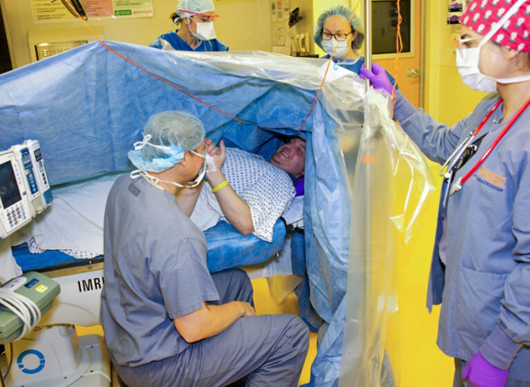
A professional musician developed a tumor in the part of the brain associated with auditory processing and could not longer differentiate musical pitches. During an awake craniotomy at UVA Health, the surgical team carefully mapped and preserved this region while removing the tumor.
“The patient regained the ability to perceive melodies and distinguish subtle pitch differences, allowing them to resume playing their instrument and enjoy their passion for music,” says neurosurgeon Melike Mut Askun, MD, PhD. “This restoration of function not only improved their quality of life but also reinstated their confidence and sense of identity.”
With a commitment to innovation and patient-centered care, UVA Health consistently delivers excellent outcomes — for this patient and others with complex neurological conditions.
Greater Surgical Precision Improves Outcomes
The team most commonly uses awake craniotomy for gliomas and epilepsy. During surgery, the team uses advanced neuromonitoring and mapping techniques, such as real-time cortical/subcortical stimulation, ECoG, neuronavigation, and electrophysiological monitoring. Patients can actively communicate with the surgical team, perform multiple tasks related to motor, language, speech and cognition. That provides surgeons with immediate feedback on the effects of surgical manipulation.
“Awake craniotomy is ideal for complex cases where standard imaging cannot fully delineate functional areas,” Mut Askun explains. “We can make intraoperative adjustments based on patient responses, enhancing the precision of the procedure.”
This approach dramatically lowers the risk of postoperative neurological deficits, including cognitive, speech, and motor impairments, compared to traditional craniotomy.
For patients with epilepsy, the team uses electrical stimulation to identify and map seizure foci and their proximity to critical functional areas intraoperatively. This mapping allows precise removal of epileptogenic tissue while avoiding damage to vital functional regions.
Preserving brain function during surgery reduces patients’ need for postoperative rehabilitation, allowing them to return to normal activities and regain independence sooner. It also significantly improves quality of life over the long term.
Elements of a Successful Awake Craniotomy Program
UVA Health’s awake craniotomy program is distinguished by its integration of patient-centered care, cutting-edge technology, and multidisciplinary collaboration. These features position it as one of the most advanced programs in the nation, contributing to both clinical excellence and neuroscience research.
Patient-Centered, Longitudinal Care
The team carefully tailors every stage of the patient journey — from preoperative planning to postoperative rehabilitation — to meet the patient’s individual needs. The program also places a strong emphasis on longitudinal follow-up. This comprehensive framework prioritizes the patient’s emotional and functional well-being.
“We customize every aspect of the procedure, from imaging and neuromonitoring to anesthesia and even finding the most comfortable surgical positioning for the patient and the team. While we follow established standards, our team’s expertise and experience allow us to adapt each step to suit the unique requirements of the patient,” Mut Askun shares.
Working closely with the neuromonitoring team, we select the most appropriate battery of tests for each patient based on their unique neurological and functional profiles. Real-time feedback during surgery allows the team to map cortical and subcortical pathways dynamically, ensuring critical functions are preserved.
Multidisciplinary Collaboration
“The success of an awake craniotomy begins with an extensive patient selection process,” Mut Askun explains.
Each candidate undergoes a thorough evaluation with our neurosurgery, anesthesia, neurology, and neuropsychology teams. This collaborative approach ensures that the decision to proceed is based not only on neurosurgical feasibility but also on anesthesia safety and the patient’s ability to actively participate during surgery.
Following that decision, patients meet with the entire multidisciplinary team. Neuropsychologists perform cognitive and functional assessments, and anesthesiologists evaluate sedation strategies and safety measures.
“This holistic preparation helps to address both the technical and emotional aspects of surgery, making patients feel supported and confident in every step of their journey,” Mut Askun says.
During surgery, our anesthesia team also plays a crucial role in maintaining optimal sedation levels while ensuring the patient remains comfortable and responsive.
Improving Techniques & Exploring New Applications
To build on its success, the UVA Health team is working to:
- Integrate advanced imaging and neuromonitoring to further refine brain mapping
- Leverage artificial intelligence to predict patient outcomes
- Enhance minimally invasive approaches
- Expand clinical trials to explore new applications for awake surgery, such as treating cognitive dysfunction in glioma patients